About clinic
The Munich Radiology Center (Conradia) provides a wide range of medical services in the fields of radiology, nuclear medicine, cardiology and therapy. The clinic employs world-class professionals who have achieved significant results in the field of preventive medicine. Their knowledge and skills contribute to the unmistakable diagnosis of patients' health, the ability to use it to map personal risks and predispositions of clients to certain diseases. Individual examination programs and doctor's recommendations not only help to improve the well-being of patients at the Munich Radiology Center, but also reduce the risk of recurrence.
The strategy of the Conradia Radiology Center (formerly Diagnostik München) is based on the prevention of the risk of disability, the maintenance of vitality and a high emotional background in patients.
All branches of the medicine in clinic
Clinic doctors
Prices for procedures
Diagnostics
Treatment
The Munich Radiology Center (Conradia) consists of seven locations, six located in Munich and one at the Munich-Planegg Urology Hospital in the suburbs.
- Significant array of survey data and extensive practical experience in the diagnosis of complex diseases.
- Large range of health diagnostic services:
- Magnetic resonance imaging with high resolution, without irradiation and dynamic layer-by-layer images.
- High resolution computed tomography, non-overlapping layer-by-layer images and low beam load.
- X-rays using the latest digital equipment that takes pictures without exposing a person to radiation exposure.
- Sonography (US) is a completely safe device that is used to obtain reliable data on the condition of the patient's organs.
- Interdisciplinary approach to diagnosis and prevention of diseases.
- A team of highly recognized doctors recognized in the medical community.
- Provision of medical care in a single set of services - diagnostics and medical examination.
- Centralized appointment.
- Staff of translators.
Magnetic resonance imaging
(MRI) is a method of diagnosis in which the patient is not irradiated during the procedure, and images are obtained using electromagnets and radio waves. This type of diagnosis is completely safe and is prescribed to children and women who are carrying a child (from the 2nd trimester).
The Munich Radiology Center has 3 "semi-open" Siemens Aera MRI machines (1.5 Tesla), 1 Siemens Skyra MRI machine (3 Tesla), a Philips Ingenia MRI machine (3 Tesla) and a Philips MRI machine (1.5 Tesla). ). Research with this equipment is especially convenient for people suffering from fear of confined spaces.
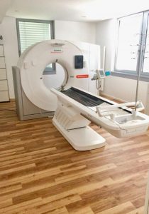
Computed tomography
CT is a radiological imaging method often used in examinations, which is used to obtain images of body parts in cross section.
In the process of research, a person is in an X-ray tube emitting X-rays. A set of detectors on the opposite side measures the radiation parameters and analyzes them. In this way, get quality pictures of internal organs and body parts.
Nuclear medicine
Based on the advanced achievements in the field of nuclear medicine, the specialists of the center seek to obtain detailed information about the state of the studied organs. Doctors obtain data on the functional capabilities of the body by administering radioactive isotopes to the patient, in addition, some of them are used in medical therapy.
As a rule, the artificially derived radioisotope technetium-99m is used in this branch of the Conradia Center in Munich. The drug is intravenous, half of its radioactive nuclei disintegrate in six hours, so it is considered fast-acting.
Digital radiography
The procedure of digital radiography is in demand in the medical research environment due to the speed and reliability of the information obtained with its help about human health.
Radiography is the production of static images of the skeleton and internal organs. It is important to note that the process does not bring discomfort to the patient and passes quickly. With its help diagnose the condition of the chest, genitourinary system, bone skeleton.
Ultrasound scanning
Ultrasound - visualization of human organs with the use of ultrasonic waves. The 2-dimensional image is displayed on the computer screen in real mode. The obtained data convey an actual picture of the state of the structure, size and shape of the research area.
This type of diagnosis is ideal for boneless areas of the body, so it is prescribed to check the soft tissues of the neck, thyroid and internal organs, in cancer diagnosis.
- Translation services.
- A shuttle service is available.
- Radiology.
- Nuclear medicine.
- Cardiology.
- Therapy.
- Urology.
- Oncology.
- Diseases of the musculoskeletal system.
- Heart disease.
- Prostatitis.
- Check-up.
- MRI.
- CT.
- Ultrasound.


